Preclinical Image Gallery
The Power of Permanent Magnet Technology
View our image gallery and see for yourself how Aspect Imaging’s compact MRI systems compare to conventional high-field superconducting MRI systems, while delivering high-resolution, quality MR imaging more easily and more
affordably for a full range of applications.
Aspect Imaging Enhancing 3D Optical Tomography through Compact MRI
M-Series™ In Vivo Applications
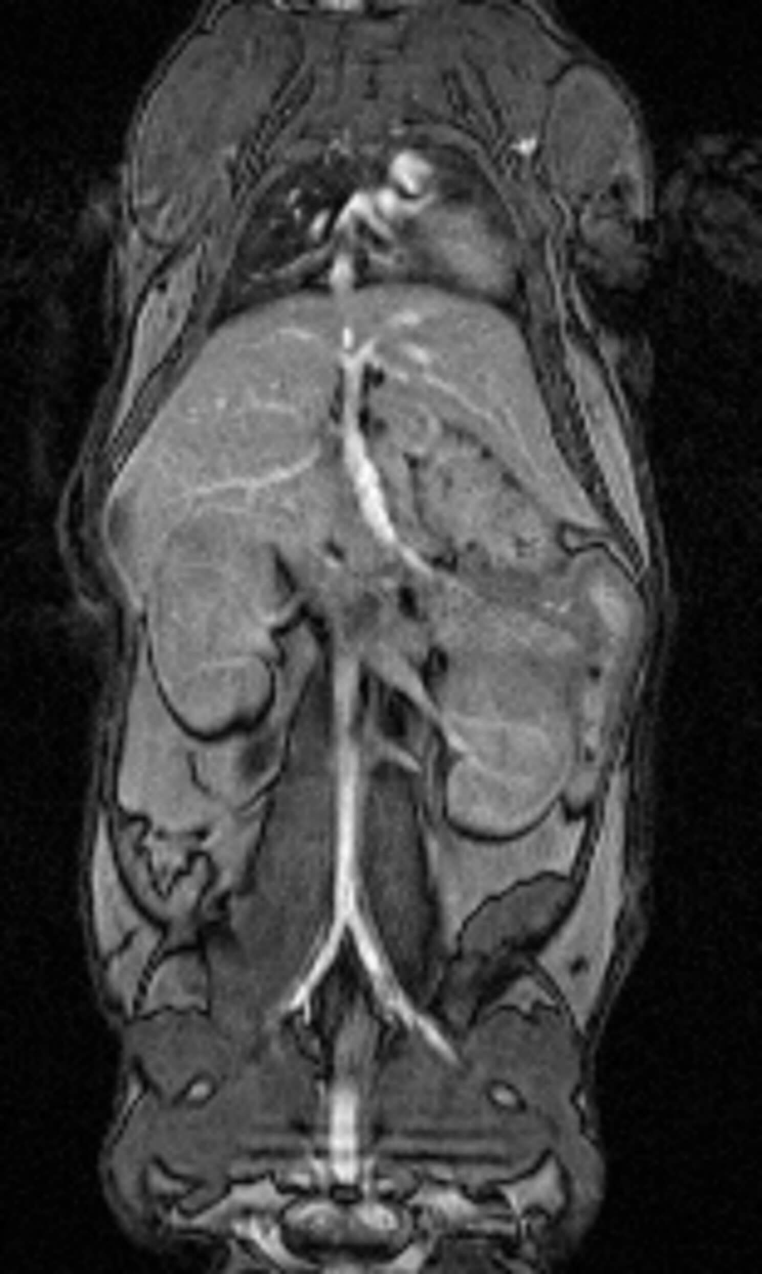
3D Whole Body Anatomy of a Normal Mouse
Fat Imaging 3D Fat Volume Quantification in a
Normal Mouse
Tumor Visualization Transgenic Pancreatic Tumor Model
Lumiquant Multi-Modal Compact MR-Luminescence Imaging of Orthotopic Metastatic Ovarian Tumor Model
Embryo Imaging — Developmental Imaging in a Normal Pregnant Mouse
Tumor Visualization and Volume Quantification — Transgenic Pancreatic Tumor Model
Cardiac Imaging of a Mouse

Mouse Head (In Vivo)
3D MR-Based Histology System
3D Visualization of Adult Rat Brain
3D REndering of E17 5 Mouse Embryo
3D MR-Based Histology Ex Vivo Rabbit Knee
Multiple Mouse Liver Focal Fatty Changes (FFC)
Histopathology of Hepatocellular Carcinoma (HCC)
3D MR-Based Histology Ex Vivo Rat Embryo E20
Fibrotic Rat Lung Volume Quantification
Mouse Brain Tumor MRI vs. Histology
Neuroscience Head Imaging – Examples
Longitudinal Evaluation of Tumor Growth In Vivo
Rat Head (Ex Vivo MRI)
Normal Rat Brain (Ex Vivo MRI)
Rat Brain (In Vivo MRI)
Rat Brain Stroke (Ex Vivo MRI)
Enhancing Visibility with Gd-Based Contrast Agents
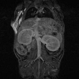
Gd-Based Contrast Agent – Heat, Vessels, Kidney, Bladder

Enhancement of Blood Vessels

Retention in Kidneys

Enhancement of Placentas in Pregnant Mouse
MRI Simplified for Preclinical Research
Our preclinical and medical imaging solutions are used by leading research
and healthcare organizations around the world.
Schedule a Virtual Demo Today!
Speak with an Aspect Imaging specialist to learn how you can benefit from the power of our patented permanent magnet technology and generate quality images with confidence.
